Skin Cancer
Comprehensive Skin Cancer Care in Southwest Missouri
A skin cancer diagnosis can feel overwhelming, but effective treatment is available right here in Springfield, MO. At Swann Dermatology, our board-certified dermatologists specialize in treating all types of skin cancer — basal cell carcinoma, squamous cell carcinoma, and melanoma — using the most advanced techniques available. We offer Mohs micrographic surgery with the highest cure rates, non-surgical brachytherapy, and photodynamic therapy for precancerous lesions. Our comprehensive approach includes prevention strategies, medical-grade sunscreens, and cosmeceuticals recommended by Dr. Swann to reduce your risk of future skin cancers. With locations throughout Southwest Missouri, we’re here to provide expert, compassionate skin cancer care close to home.
Our Skin Cancer Treatment Options

Mohs Micrographic Surgery
During Mohs surgery, our fellowship-trained surgeons remove thin layers of cancerous tissue one at a time, examining each layer immediately under a microscope until no cancer cells remain. This real-time verification means you leave with your cancer completely removed and your wound reconstructed in a single 2-5 hour visit — no waiting for lab results, no additional surgery. Our Springfield Mohs surgery specialists have performed over 20,000 Mohs procedures and bring the highest level of expertise in skin cancer removal and reconstruction to patients across Southwest Missouri. At this time, we are performing Mohs surgery at three of our clinics: Springfield, Hollister, and Lebanon.

Brachytherapy | A Radiation Alternative to Mohs
Brachytherapy is different from traditional external beam radiation therapy. With brachytherapy, the dose-curves are steep, which means that the radiation field is kept very tight and small. Cure rates are not as high as Mohs surgery but approach 95-97% without surgery. Esteya is a patient-friendly, electronic brachytherapy solution for treating skin cancer. Specifically designed for non-melanoma skin cancer treatments, Esteya provides a non-surgical treatment that destroys cancer cells while sparing healthy surrounding tissue. There’s minimal or no recovery period — which means there will be negligible impact on your daily activities.

Photodynamic Therapy
Photodynamic therapy (PDT) treats precancerous actinic keratoses using a light-activated medication applied directly to sun-damaged skin. We apply aminolevulinic acid (ALA) to the treatment area, then activate it with blue or red light to destroy precancerous cells while preserving healthy tissue. This in-office procedure is ideal for patients with numerous precancers — typically 15 or more — on the face, scalp, or neck, especially those with extensive sun damage who are at high risk for developing skin cancer. A series of PDT treatments can significantly reduce existing precancers and help prevent new ones from forming.
Understanding Skin Cancer: Types, Detection & Prevention
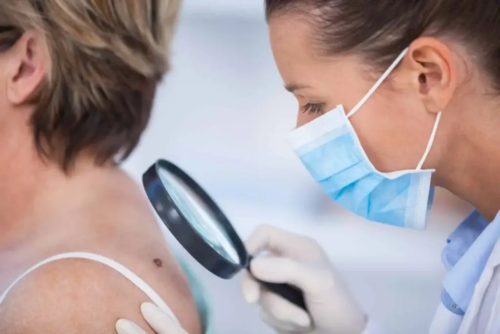
Skin cancer is the most common cancer in the United States, with more than a million new cases diagnosed annually. One in five Americans will develop skin cancer during their lifetime, but when caught early, cure rates are excellent. At Swann Dermatology, we specialize in comprehensive skin cancer management—from early detection and screening to advanced treatment options including Mohs surgery, brachytherapy, and photodynamic therapy.
- Fair skin or light-colored eyes
- History of sunburns or excessive sun exposure
- Active outdoor lifestyle without adequate sun protection
- Family history of skin cancer
- Age over 50
- Previous skin cancer diagnosis (increases risk of developing new cancers)
- Basal Cell Carcinoma – The most common type, accounting for about 70% of cases. Slow-growing and rarely spreads, but requires treatment.
- Squamous Cell Carcinoma – The second most common, making up about 20% of cases. More aggressive than basal cell and can spread if left untreated.
- Melanoma – The least common but most dangerous form. Can spread rapidly to internal organs and the lymph system, making early detection critical.
Approximately 90% of basal and squamous cell carcinomas are caused by overexposure to ultraviolet (UV) light, making prevention and early detection your best defense. Regular self-examinations and annual skin screenings with a dermatologist are essential, especially if you’ve had skin cancer before. When examining your skin, look for changes in:
- Size
- Shape
- Color
- Diameter
- Texture or elevation
If you notice any suspicious changes, contact us immediately. Early-stage skin cancers are highly treatable, with excellent cure rates when diagnosed promptly.
Types of Skin Cancer
Basal Cell Carcinoma
This is the most common form of skin cancer. Basal cells reside in the deepest layer of the epidermis, along with hair follicles and sweat ducts. When a person is overexposed to UVB radiation, it damages the body’s natural repair system, which causes basal cell carcinomas to grow. These tend to be slow-growing tumors and rarely metastasize (i.e., spread). Basal cell carcinomas can present in a number of different ways:
- Raised pink or pearly white bump with a pearly edge and small, visible blood vessels
- Pigmented bumps that look like moles with a pearly edge
- A sore that continuously heals and re-opens
- Flat scaly scar with a waxy appearance and blurred edges
Despite the different appearances of the cancer, they all tend to bleed with little or no cause. Approximately 85 percent of basal cell carcinomas occur on the face and neck since these are areas that are most exposed to the sun.
Risk factors for basal cell carcinoma include fair skin, sun exposure, age (most skin cancers occur after age 50), exposure to ultraviolet radiation (from tanning beds, for example), and therapeutic radiation given to treat an unrelated health issue.
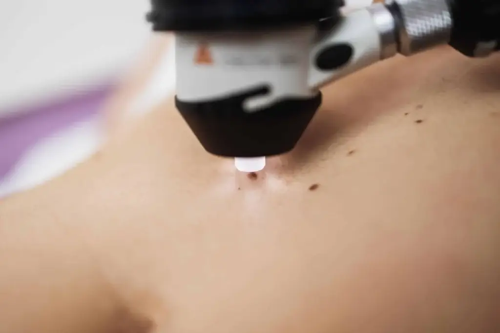
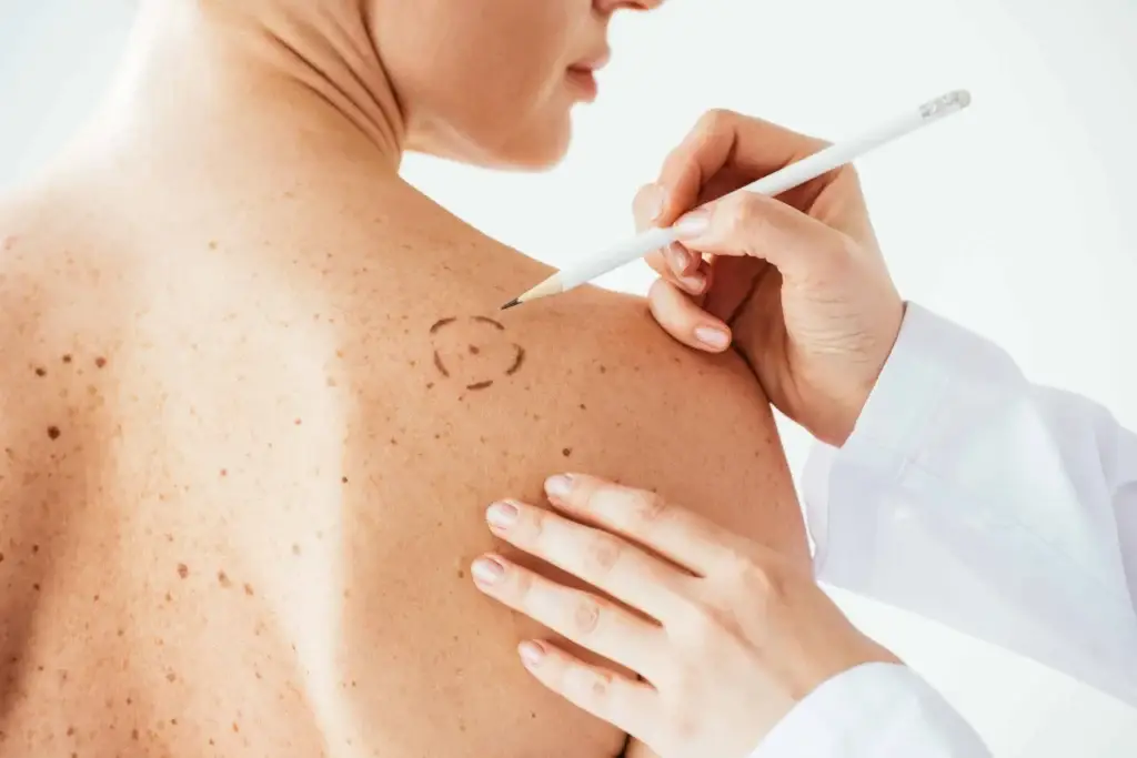
Diagnosing basal cell carcinoma requires a biopsy — either excisional, where the entire tumor is removed along with some of the surrounding tissue, or incisional, where only a part of the tumor is removed (used primarily for large lesions).
Treatments for Basal Cell Carcinoma
At our Springfield skin cancer clinic, we offer multiple treatment options for basal cell carcinoma:
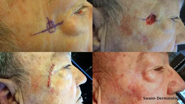
- Mohs Micrographic Surgery — Mohs is the preferred treatment method for optimal management of complex tumors, including those on the head and neck, large tumors, or tumors previously treated by other methods where recurrence of the cancer has been detected. Mohs Micrographic Surgery combines removal of cancerous tissue with microscopic review while the surgery takes place. By mapping the diseased tissue layer by layer, less healthy skin is damaged when removing the tumor.
- Cryosurgery — Some basal cell carcinomas respond to cryosurgery, where liquid nitrogen is used to freeze off the tumor.
- Curettage and Desiccation — The preferred method of dermatologists, this treatment involves using a small metal instrument (called a curette) to scrape out the tumor, followed by an electric current to destroy any remaining cancer cells and seal the wound. This method is ideal for small, well-defined basal cell carcinomas on low-risk areas of the body where the cosmetic outcome is less critical.
- Prescription Medicated Creams — These creams can be applied at home. They stimulate the body’s natural immune system over the course of weeks.
- Radiation Therapy — Radiation therapy is used for difficult-to-treat tumors, either because of their location, severity or persistence.
- Surgical Excision — The tumor is surgically removed and stitched up.
Squamous Cell Carcinoma
Squamous cells are found in the upper layer (the surface) of the epidermis. They look like fish scales under a microscope and present as a crusted or scaly patch of skin with an inflamed, red base. They are often tender to the touch. It is estimated that 250,000 new cases of squamous cell carcinoma are diagnosed annually, and that 2,500 of them result in death.
Squamous cell carcinoma can develop anywhere, including inside the mouth and on the genitalia. They most frequently appear on the scalp, face, ears and back of hands. Squamous cell carcinoma tends to develop among fair-skinned, middle-aged and elderly people who have a history of sun exposure. In some cases, it evolves from actinic keratoses, dry scaly lesions that can be flesh-colored, reddish-brown or yellow black, and which appear on skin that is rough or leathery. Actinic keratoses spots are considered to be precancerous.
Like basal cell carcinoma, squamous cell carcinoma is diagnosed via a biopsy — either excisional, where the entire tumor is removed along with some of the surrounding tissue, or incisional, where only a part of the tumor is removed (used primarily for large lesions). Our Springfield dermatologists use advanced diagnostic techniques to accurately stage squamous cell carcinoma and recommend the most effective treatment.
Treatments for Squamous Cell Carcinoma
- Mohs Micrographic Surgery — Since squamous cell carcinomas can be more aggressive than basal cell carcinomas, Mohs surgery is often the best treatment option because it has the highest cure rate. Mohs Micrographic Surgery combines removal of cancerous tissue with microscopic review while the surgery takes place. By mapping the diseased tissue layer by layer, less healthy skin is damaged when removing the tumor.
- Surgical Excision — With this treatment option, the tumor is surgically removed and stitched up. The tissue is sent in a jar to a pathologist, who sections the tissue and makes slides, then reads the slides to determine if the representative margins appear clear. It takes approximately 5 business days to determine if the margins are clear or if further surgery is warranted.
- Radiation Therapy — Traditional radiation therapy is used for difficult-to-treat tumors, either because of their location, severity or persistence. Electronic brachytherapy can be utilized in-office to treat relatively early primary tumors with a high cure rate and no surgical scar.
Melanoma
While melanoma is the least common type of skin cancer, accounting for only 1% of cases, it is by far the most deadly. This aggressive cancer can spread rapidly to internal organs and the lymph system, making early detection absolutely critical. Melanoma is also the most common form of cancer among young adults ages 25 to 29.
Melanoma develops in melanocytes, the cells that produce melanin and give skin its pigmentation. Because of this, melanomas typically appear as dark brown or black spots on the skin. They often resemble moles or grow inside existing moles, which is why regular self-examinations are essential for early detection. When caught in its earliest stages, melanoma is highly curable, but once it spreads, treatment becomes significantly more challenging. At Swann Dermatology in Springfield, we emphasize early melanoma detection through comprehensive skin cancer screenings and patient education.
The primary cause of melanoma is overexposure to the sun, especially severe sunburns during childhood. This cancer also tends to run in families, so those with a family history should be particularly vigilant about monitoring their skin. Other risk factors include fair skin, light-colored eyes, and having many moles.
Diagnosis requires a biopsy to examine the suspicious tissue and determine the depth and stage of the cancer. Treatment options range from surgical removal for early-stage melanomas to immunotherapy, targeted therapy, radiation, or chemotherapy for more advanced disease. Early detection and prompt treatment offer the best chance for cure.
How to Spot Melanoma
The key to detecting skin cancers is to notice changes in your skin:
- Large brown spots with darker speckles located anywhere on the body
- Dark lesions on the palms of the hands and soles of the feet, fingertips, toes, mouth, nose or genitalia
- Translucent pearly and dome-shaped growths
- Existing moles that begin to grow, itch or bleed
- Brown or black streaks under the nails
- A sore that repeatedly heals and re-opens
- Clusters of slow-growing scaly lesions that are pink or red
- Asymmetry — Half the mole does not match the other half in size, shape or color.
- Border — The edges of the mole are irregular or blurred.
- Color — The mole is not the same color throughout.
- Diameter — The mole is larger than one-quarter inch in size.
- Elevation — The mole becomes elevated or raised from the skin.
If you notice any of these warning signs, please make an appointment to see one of our dermatologists right away. The doctor may do a biopsy of the mole to determine if it is or isn’t cancerous.
How to Reduce Your Risk of Skin Cancer
Since 90% of basal cell and squamous cell carcinomas are caused by UV exposure, protecting your skin from the sun is your best defense:
- Stay out of the sun during peak hours, between 10 a.m. and 4 p.m.
- Cover your arms and legs with protective clothing.
- Wear a wide-brimmed hat and sunglasses.
- Use sunscreens with an SPF of 15 or greater year-round, and make sure they work on both UVA and UVB rays. Look for products that use the term “broad spectrum.”
- Check your skin monthly, and contact your dermatologist if you notice any changes.
- Schedule regular skin examinations. Adults over 40 should have annual skin exams with a dermatologist.
Schedule Your Skin Cancer Consultation in Springfield, MO
If you’re concerned about a suspicious mole or lesion, don’t wait. We serve patients throughout Southwest Missouri, including Springfield, Nixa, Ozark, Republic, and Branson. Our experienced skin cancer doctors offer:
- Same-day and next-day appointments
- On-site biopsies and pathology
- Comprehensive skin cancer screenings
- All treatment options under one roof