Skin Cancer Screening
Full-Body Skin Exams in Springfield, Branson, & Southwest Missouri
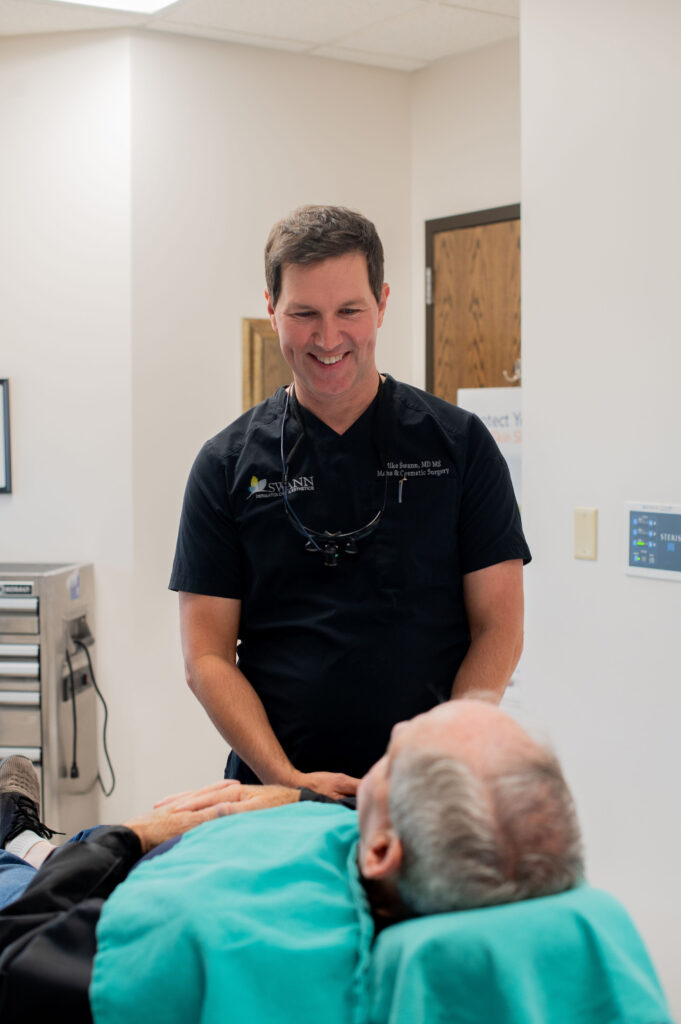
What to Expect During Your Skin Exam
A skin cancer screening is one of the simplest appointments you’ll have with us — and one of the most important. Here’s exactly how it works, so nothing about it is a surprise.
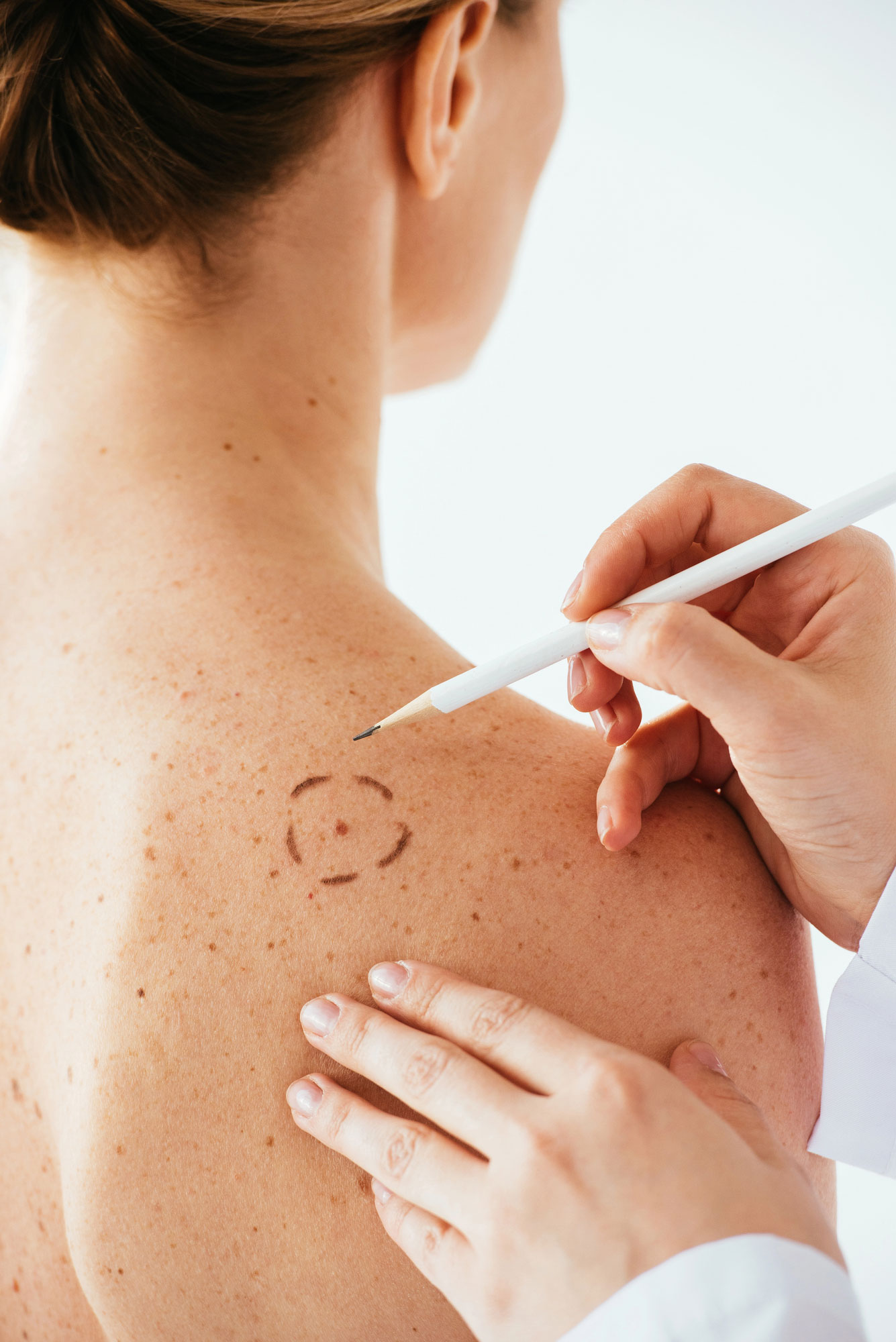
When you arrive, you’ll change into a gown so your dermatologist can examine your skin from head to toe. Using a bright light and a handheld magnifier called a dermatoscope, your provider checks the places you’d expect — your face, arms, chest, back, and legs — as well as the spots that are easy to miss on your own: your scalp, behind your ears, between your fingers and toes, the soles of your feet, and under your nails. The exam is thorough but quick, usually taking about 10 to 15 minutes, and it’s completely painless.
As we go, we’re looking for anything that stands out: a mole that has changed, a spot that looks different from your others, or a sore that won’t heal. If we see something that needs a closer look, we will talk you through it and, in most cases, can perform a simple in-office biopsy the same day rather than asking you to come back. You’ll never leave wondering what we saw — we explain what we find and what, if anything, happens next.
How to Prepare for Your Appointment
- Remove nail polish from both fingers and toes — skin cancers can develop under the nails, and we need to see them clearly.
- Come with clean skin and minimal makeup so we can examine your face fully.
- Wear your hair loose rather than in a tight style, so we can check your scalp.
- Be ready to remove jewelry if needed.
- Make a note of any spots that concern you — a mole that's new, changing, itching, or bleeding — so we can be sure to look at them.
- Know your history. If you or a close relative has had skin cancer, or if you've used tanning beds or had significant sun exposure, let us know so we can tailor the exam to you.
How Often Should You Be Screened?
For most adults, a professional skin exam once a year is the right rhythm. Skin cancer is highly treatable when it’s caught early, and an annual check is the most reliable way to find something while it’s small.
Some people benefit from more frequent screenings. Your dermatologist may recommend coming in every six months — or sooner — if you have any of the following:
- A personal history of skin cancer or precancerous lesions
- A family history of melanoma or other skin cancers
- Fair skin, light eyes, or skin that burns easily
- A large number of moles, or any unusual or atypical moles
- A history of significant sun exposure, sunburns, or tanning bed use
- A weakened immune system
Between visits, it’s worth checking your own skin about once a month (see below) and booking an appointment any time you notice a spot that’s new, changing, or won’t heal — you don’t have to wait for your annual exam.
How to Check Your Own Skin (the ABCDEs)
Monthly self-exams are one of the best ways to catch a problem early, because you’re the person most likely to notice when something on your skin changes. Pick a well-lit room, use a full-length mirror and a hand mirror for the spots you can’t see directly, and don’t forget your scalp, the soles of your feet, between your toes, and under your nails.
When you’re looking at a mole or spot, the American Academy of Dermatology’s ABCDE guide is an easy way to know what’s worth a closer look:
- A — Asymmetry: One half of the mole doesn't match the other.
- B — Border: The edges are irregular, ragged, or blurred.
- C — Color: The color isn't uniform, or includes shades of brown, black, red, white, or blue.
- D — Diameter: The spot is larger than about a quarter inch (the size of a pencil eraser), though melanomas can be smaller.
- E — Evolving: The mole is changing in size, shape, color, or how it feels — or it's new.
If a spot checks any of these boxes, don’t wait for your next scheduled visit. Contact us and we’ll take a look.
If We Find Something
If your screening turns up a suspicious spot, we’ll confirm with a biopsy and walk you through the right treatment for it — whether that’s Mohs micrographic surgery for the highest cure rate, non-surgical electronic brachytherapy, or photodynamic therapy for precancerous lesions. Whatever the path, you won’t navigate it alone. We handle every step of the process — screening, diagnosis, and treatment — under one roof.
Why Choose Swann Dermatology
When the topic is skin cancer, experience matters. At Swann Dermatology, your screening is performed by board-certified dermatologists who diagnose and treat skin cancer every day — and if you ever need treatment, you’re already in the hands of one of the most experienced teams in Southwest Missouri. Our fellowship-trained Mohs surgeons have performed more than 20,000 Mohs procedures, and because we offer the full range of skin cancer treatments under one roof — Mohs surgery, brachytherapy, and photodynamic therapy — you’re never sent elsewhere to finish your care.
Schedule Your Skin Cancer Screening in Southwest Missouri
Early detection starts with a single appointment. We provide skin cancer screenings for patients across Southwest Missouri, including Springfield, Branson, Lebanon, and Monett. If you’ve noticed a spot that worries you, or it’s simply been more than a year since your last skin check, now is the time.
FAQ's
Does a skin cancer screening hurt?
How long does a skin exam take?
Do I have to undress completely?
At what age should I start getting screened?
What's the difference between a skin cancer screening and a mole check?
What happens if you find something suspicious?
What happens if you find something suspicious? We’ll explain what we see and, in most cases, can perform a simple biopsy the same day. If it turns out to be skin cancer, we’ll guide you through your treatment options right here in our practice.